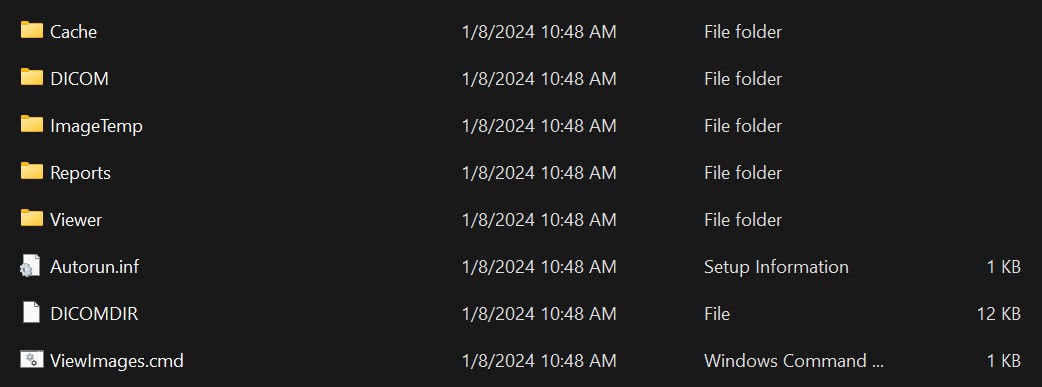
I took the CD of my Liver Ultrasound over to Daniels last night and saved the contents to a thumb drive. I looked at the files this morning and there was no viewer software for the DICOM image file format included. Which means it's totally unusable to a layman. Only people in the Medical profession have the software to view these images.
Unless you're a computer genius like me, who can track down a Dicom viewer package and install it on my Windows 11 laptop.
Bingo, there were 37 images to do with as I wanted. I could have converted them all to .jpg, but the viewer app had an export to video function, so I created one.
The resulting video had my confidential info on the front and back, and it flew through the 37 images in three seconds. So, I popped it into my video editor, stripped off the personal data, and stretched it out:
The video turned out great. You can see the title of the organ, force it's way into the upper left, as each one slides into view.
Here is the good doctor Douglas White's diagnosis as a result of seeing these images.
• Pancreas is unremarkable.
• Gallbladder is normal.
• Common Bile Duct is good.
• Right Kidney has mild diffuse renal cortical thinning without hydronephrosos or nephrolithiasis.
Bottom line, he stated that I have diffuse fatty infiltration of the liver, otherwise unremarkable.
Sometimes you have to dive deeper when all they say is "You have a fatty liver, so now fuck off, that's all you get to know".
